New research uncovers crucial differences in tumor environments for people with folliculotropic mycosis fungoides compared to the classic form.
Recent advances in medical imaging technology are shedding new light on a rare skin condition that can affect people with albinism and the general population alike.
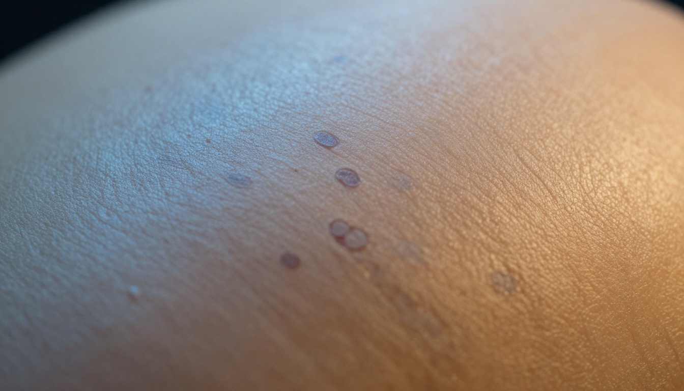
Researchers using spatial transcriptomics have discovered significant differences in the tumor environments of two forms of mycosis fungoides (MF), the most common subtype of cutaneous T-cell lymphoma. The study, published in the Journal of Investigative Dermatology, compared folliculotropic mycosis fungoides (FMF) with the classic form of the disease.
According to the research, mycosis fungoides is characterized by the proliferation of malignant T cells in the skin. The study reveals that in the more aggressive folliculotropic variant, these malignant cells specifically target hair follicle structures, while in classic MF, they spread throughout the dermis and epidermis layers of the skin.
This distinction in cell behavior may help explain why folliculotropic mycosis fungoides tends to be more clinically aggressive. The researchers utilized advanced spatial transcriptomics technology to analyze patient samples and identify these crucial microenvironmental differences between the two forms.
For the albinism community, this research carries particular significance. People with albinism already face increased skin sensitivity and higher risk for certain skin conditions due to reduced melanin protection. Understanding the subtle differences between skin lymphoma variants could potentially lead to earlier detection and more targeted treatments.
Though rare, mycosis fungoides represents one of many skin conditions that healthcare providers should be vigilant about when caring for patients with albinism during regular skin checks.
Keywords
Core topics and entities mentioned in this summary.